Posts

Showing posts from August, 2013

The Rotator Cuff

Image
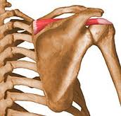
What is the rotator cuff? A group of muscles surrounding the shoulder joint: Supraspinatus: Location: Supraspinous fossa of scapula (shoulder blade) to greater tubercle of humerus (upper arm bone). Basic action: Assists deltoid in abduction (out to the side movement) of the humerus. Infraspinatus: Location: Infraspinous fossa of the scapula to greater tubercle of the humerus. Basic action: Externally rotates (turns out) the humerus. Teres Minor: Location: Lateral border of the scapula to greater tubercle of the humerus. Basic action: Externally rotates the humerus. Subscapularis: Location: Subscapular fossa of the scapula to lesser tubercle of the humerus Basic action: Internally rotates (turns in) the humerus. However these basic actions don’t occur in function as the cuff work together to do something very different! So what does the rotator cuff actually do? Aids movement of the glenohumeral joint (shoulder joint) Comp
The Vastus Medialis Oblique (VMO)

Image
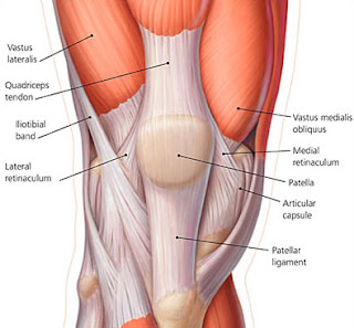
Despite the emphasis placed upon the importance of VMO training to re-balance the quadriceps in patellofemoral pain syndrome, it's still relatively unknown whether the timing of Vastus Medialis Oblique (VMO) & Vastus Lateralis (VL) contraction is a risk factor for patellofemoral pain syndrome (knee cap pain).   To add to this issue, debate exists as to whether the Vastus Medialis oblique (VMO) can be activated to a greater degree than the Vastus Lateralis to produce preferential strengthening. What does the evidence say? For: In closed kinetic chain exercises, more selective VMO activation can be obtained at 60° knee flexion (Tang et al 2001). Performing quads exercises with EMG biofeedback improved the muscle activity balance between VMO & VL (Ng et al 2008). Lunges produced the VMO:VL ratio (1.18:1), which is the closest to the idealized ratio of 1:1 (Irish et al 2010). Corrective taping is effective in reducing pain in patellofemoral p

Muscle blog series: Part 8: Core stability:

Image
This is the next blog article in the series and will discuss core stability. What is Core Stability? Core stability is defined as the ability to control the position and motion of the trunk over the pelvis to allow optimum production, transfer and control of force and motion to the terminal segment in integrated athletic activities. Core muscle activity is best understood as the pre-programmed integration of local, single-joint muscles and multi-joint muscles to provide stability and produce motion. This results in proximal stability for distal mobility, a proximal to distal patterning of generation of force, and the creation of interactive moments that move and protect distal joints. What is the background of core stability? Core stability started in the late 1990’s. It was derived from studies that demonstrated a change in timing of the trunk muscles in back injury and chronic lower back pain (CLBP) patients. However , there have been assumption