Patellofemoral pain syndrome: A complex issue.

Knee pain is rarely caused by isolated events, more commonly they are the consequences of habitual imbalances in the movement system. The one off injury such as the torn anterior cruciate ligament can and does occur, but these are relatively simple as we know what the injury is and what caused it, which makes it easier to treat and prevent re-occurrences.
However, physiotherapists are dealing with more complex pain
syndromes such as patellofemoral (knee
cap joint) pain, shin splints, and back pain, amongst many others. The issue
with these types of conditions is that the cause may be difficult to establish
and may be many things. Even more complicated the pain may be coming from
another area of the body and not from the site of pain! Equally it may be
caused in part by the postures and activities that are performed every day,
which could be work related. So the problem then becomes how to you recover and
prevent when you are constantly causing the problem every day?
Why are some individuals more susceptible to pain?
Stability:
An individual’s mechanics has a marked effect on his/her
inherent stability and passive control and hence propensity for experiencing
pain. A joint can be passively unstable, but dynamically stable, as muscles via
the neural system can compensate for the lack of stability in the passive
structures (Panjabi, 1992).
Equally, a passively stable joint can be dynamically
unstable when muscle control is poor.
Overload:
The amount of load through the soft tissues or the frequency
of the loading will also affect joint structures and may result in tissue failure,
as an individual may breach his/her threshold and stray out of the zone of
homeostasis ( Dye, 1996). Dye contends that symptoms will not be present if an
individual is operating inside his/her envelope of function, but as soon as a
threshold is reached a complex biological cascade of trauma and repair will
occur, which will be manifested clinically by pain and swelling.
What factors can contribute to patellofemoral pain?
The shape of your bones can actually affect the knee cap
position and movement due to effects on muscle activation, for example: at the
hip, femoral anteversion or internal femoral rotation affects the position of the
trochlea relative to the patella. Internal femoral rotation is associated with
poor posterior gluteus medius activation, affecting the stability of the
pelvis. So these effects are not just confined to the knee. A prime example of
secondary effects is back pain.
So in this example what can help?

Improving hip extension and external rotation mobility as well
as gluteal control in weight bearing by simulating the stance phase of gait,
can significantly improve the symptoms of many “difficult” knees.
Other potential causes:

Pronation problems of the foot – too much, too long or too
late can cause an increase in the dynamic valgus vector force on the
patellofemoral joint, as this will cause a chain reaction of events up the
body.
So if I pronate, what can I do?

Orthotics may be used to help foot control. Clinically, comfort
is the most important and relevant feature when prescribing foot orthoses
(Mündermann et al 2003).
What about causes directly related to the knee?

Locally, tightness of the lateral retinacular structures and
the iliotibial band as well as delayed onset of the Vastus Medialis Oblique (VMO)
relative to the Vastus Lateralis (VL) can cause pressure distribution variations
of the patella on the femur. Changing the activity of the VMO relative to the
VL may not only require up-training of the VMO but may require downtraining the
VL. Some “difficult” knees may require inhibitory tape on the VL to improve the
coordination of the quadriceps activation.
Where is the pain coming from?
It’s not as simple as you might think there can be multiple
local anatomical structures causing the
knee pain. Those implicated include the medial and lateral retinaculum,
the patellar subchondral bone, the anterior synovium, the joint capsule, the
patellar tendon, the infrapatellar fat pad and scarring of the infrapatellar
branch of the saphaneous nerve following arthroscopy. The fat pad is highly
nociceptive and vascular. A recent study showed that injecting 0.25 ml
hypotonic saline into the fat pad of asymptomatic individuals caused moderate
to severe anterior knee pain (Bennell et al 2004).
What about referred pain?

There is always a distinct possibility of referred pain from
the lumbar spine or in prepubescent sufferers the possibility of a slipped
femoral ephiphysis in the hip!
What about osteoarthritis?
Causes of this pain can be:
Subarticular bone attrition
Bone marrow lesions
Synovitis/effusion
Meniscal tears
(Torres et al 2006)
Fat pad irritation

The fat pad is an under-diagnosed, poorly recognised source
of patellofemoral pain. Fat pad problems are frequently confused with patellar
tendinopathy.
What does the fat pad do?
The fat pad stabilises the patella at the extremes of knee
motion and alters knee biomechanics, improving tibial external rotation relative
to the femur (Bohnsack et al 2004).
So what causes fat pad irritation?
Imaging abnormalities seen are usually the consequence of
trauma and degeneration. The commonest traumatic lesions are often seen after
having an arthroscopy. So think carefully about going under the knife!
Fat pad impingement can also occur after patellar
dislocation (Saddik et al 2004).
How do I know if I have fat pad irritation?
Often acute onset of pain, associated with rapid extension
of knee such as a tumble turn in swimming or landing on a straight knee during
a running or jumping action. In standing, the inferior pole (base) of patella
is embedded in underlying tissues, the fat pad has a puffy appearance and there
is often tenderness of inferior pole of the patella. Individuals with fat pad
symptoms have very poor inner range quadriceps control and often walk with a
flexed knee gait to avoid irritating the fat pad, as straight knee positions
will hurt.
What not to do:
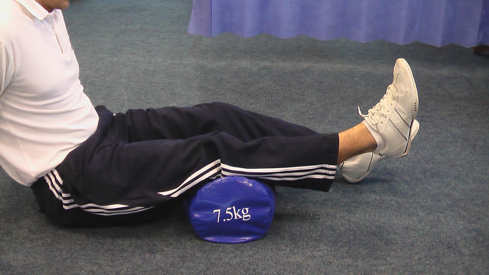
Quadriceps setting and extension overpressure manoeuvres can
exacerbate the symptoms. So kicking a football or kicking in swimming will be
painful and irritate it further.
What can be done to help?
Unless the fat pad is unloaded it is difficult for patients
to progress as the fat pad is easily irritated during daily activities and
rehabilitation.
Why doesn’t the knee heal and recover?
Inhibition:
Pain has an inhibitory effect on muscle activity and the VMO
is more affected than the VL. This creates greater muscle imbalance and
therefore more mal-tracking and therefore more pain, which in turn inhibits the
VMO more, causing more pain etc. etc. So a vicious cycle ensues.
Fear:
Fear of pain changes muscle activation patterns. Randomly
applying an electric shock across the knee during stair ascent and descent in
subjects with no history of knee pain causes a significant reduction in VMO EMG
activity but not VL activity (Hodges et al 2009). Thus, not only will pain
decrease quads activation but fear of pain will increase the imbalance between
the VMO and VL making rehabilitation difficult.
Fear-avoidance has long been recognized as an important
factor in the development of pain-related disability (Boersma et al 2005). Exposure
to stress initiates the secretion of several hormones, including
corticosterone/cortisol, catecholamines, prolactin, oxytocin, and renin, as
part of the survival mechanism. (Van de Kar et al 1999). Release of cortisol
can be detrimental to a patient’s recovery. In fact it has been found that
stress-related hormones can alter inner ear fluid homeostasis and auditory
function. (Juhn et al 1999).
Negative self talk affects a patient’s belief system can
affect outcome of treatment. Positive affirmations about the knee and
desensitising techniques can be useful in shifting the focus for the
“difficult” knee patient.
Central sensitisation:
Pain can cause permanent changes in the spinal cord, which
can then be the origin of the pain instead of the injury site, this means the
initial injury can be gone but the pain continues any way. Centrally maintained
pain can significantly affect progress and clinicians need to be aware of the
treatment options available for this group of patients as the management is
quite different (Butler & Moseley, 2003).
So what can be done to manage things when this happens?
Try to get out of end of range positions to minimise the
stress on soft tissue structures, by adapting the way that you move performing
your day to day activities.
Specific exercises which fit into daily life and can be
performed frequently, in small quantities, building for endurance, to change the
behaviour pattern (the synergistic recruitment pattern) of the muscles.
Awareness that the condition is managed not cured – so an
on-going maintenance is required. The program must be simple, requiring very
little equipment so it is transportable and takes no more than 5 minutes per
day.
I’m now recovered, does this mean I don’t have to do anything anymore?
No!
Once the knee problem has been resolved the clinician needs
to review the patient every 6 months to ensure the patient is still doing the
maintenance exercises, to keep him/her symptom free. Otherwise the problem is
likely to keep re-occurring.


in such cases physiotherapy can do wonders.
ReplyDeletephysiotherapy treatment singapore
Hi Dear,
ReplyDeleteThanks for posting this blog. I am impressed to your knee pain treatment. It is very useful for me other knee patient. Please visit at "Osteoarthritis Knee", i hope you will satisfied with us.
Visit Here - https://mens-health.sg/joint-pain-diagnosing-treatment-and-management/
Thanks Regards,,