Patella Tendinopathy

I hear different terminology used all the time, so what is tendinitis, tendinosis or tendinopathy?
Tendinopathy:
This is a broad term encompassing painful conditions
occurring in and around tendons, which includes tendinitis and tendinosis.
Tendinitis:
This basically means inflammation of a tendon.
Tendinosis:
This means degeneration of tendon (Meaning
cumulative damage).
So what does this mean to me?
For a long time tendinitis was the term used and it was
considered that injured tendons were simply inflamed. Then the thinking shifted
to the thought that there was little or no inflammation present in these
conditions (Andres & Murrell 2008). However as is with research, the
thinking is changing again: There may be an inflammatory response associated
with chronic tendinopathy, even though recent opinions had decided it to be
non-inflammatory (Rees 2013).
So both types can occur!
This is important as the way that you treat each one has to
be very different.
So how does it get injured?
Microtrauma of the patella tendon occurs when it is
subjected to extreme forces such as rapid acceleration -deceleration, jumping
& landing (Ferretti et al 2002).
It’s a case of overload,
underload or over stretch. A great example of the load that can go
through the knee below:
Knee cap force is 3 x body weight in running & 6 x body
weight in squatting & jumping (Reid 1992). There is an incredible 17 times body weight being
placed on the patellar tendon in Olympic weight lifters (Rutland et al 2010).
Bet you didn't expect that did you!
This being said though basically tendinopathy occurs from
cumulative overloading or overstretching or from lack of loading or lack of
flexibility.
So what specifically causes these effects?
Recovery time:
If you don’t allow enough recovery between
loading activities then the tendon hasn't got enough time to repair, and
training again on it too soon will build cumulative damage. A long healing time
is required for a tendon to heal due to poor vascularisation (blood flow)
(Nowak & Handyford 2000). Some people believe just to recover the
microtrauma from a run takes at least 24 hours on average to repair. So if you
do more than the average distance or heal slower than average person then you
will need more like 48 hours.
Biomechanics:

Biomechanics is the way we move and are aligned. So abnormal movements or alignments will create excessive and abnormal forces through our body. For example: Internal femoral rotation is associated with poor gluteus medius activation, affecting the stability of the pelvis and causing overpronation. Pronation of the foot: too much, too long or too late can cause an increase in the dynamic valgus vector force on the patellofemoral joint and cause patella tendinopathy. Sounds complicated but essentially abnormal movement and alignment has knock on effects through your body causing abnormal and excessive forces that can lead to injury.
Fatigue:
When the muscle is fatigued the contraction
strength is not as strong or fast, leading to more load or stretch being passed
to the tendon. Exercise can disturb proprioception through fatigue, this has
implications for musculoskeletal injuries (Proske & Gandevia 2012).
Obviously when you fatigue your movement alters, which will change the way that
your tendon is loaded. For example, when running, if your gluteus medius tires
then the pelvis will drop on the opposite side, the hip will internally rotate on
your weight bearing leg, causing overpronation in the foot. This alignment in
the foot will change the angle of load through the knee and patella tendon from
an ideal vertical direction to a lateral one. The reason that this is
significant is down to the structure of the tendon (The fibres line up
vertically and not laterally, so it is very strong in the vertical plane but
very weak in the lateral one).
Flexibility:
Another specific reason is lack of flexibility: If the Quadriceps and hamstring are too short
for the activity at hand then it is easier to potentially tear and if you don’t
warm the tissue up before activity then the tissue is not as pliable making it
tighter and more likely to damage. It was found that patients with
patellafemoral (kneecap) pain had shorter hamstrings than asymptomatic controls
(White et al 2008). Also decreased ankle dorsiflexion movement was found to be a risk factor
for developing Patella Tendinopathy in basketball players (Backman &
Danielson 2011).
Interestingly excessive flexibility is another factor:
Some people who are hypermobile (double jointed) or people
who have overlengthened their tendon are more likely to have problems as the
tensile strength of the tendon is reduced, making it easier to overload.
Muscle and tendon weakness:
If the muscle isn't strong enough and hasn't been trained to take the loads of the sport (underload cause) then it can overload and tear. This is true of tendon also.Age:
Aging & Disuse contribute to a tendon's loss of
resilience & strength (Nowak & Handyford 2000). And this is where
loading (strength training) is even more important than ever to compensate for
the loss of strength.
So what’s the chance of getting Patella tendinopathy?
There is a 22% incidence of patella tendinopathy in the athletic
population (Lian et al 2005) and
it accounts for 19% of running injuries
(Taunton et al 2002).
But if you do activities that you haven’t prepared your body
for, then you are more likely to get it.
How long does it generally last for?
Patellofemoral pain took 77 days on average to recovery in
novice runners (Nielsen et al 2014).
However if you don’t rectify the cause of your symptoms then
it will either persist or come back again in the future.
What are the symptoms of Patella tendinopathy?
Tenderness on palpation of the inferior pole of the patella
is accurate for diagnosis of patella tendinopathy (Cook et al 2001). See the
picture below:

Overall pain is brought on when ascending or descending
stairs, performing single leg declining squats, jumping or hopping (Purdam et
al 2003).
Symptoms and onset can help you to work out if it tendinosis or tendinitis:
Tendinosis:
Tends to be in the older age group and usually
comes on for no reason (no overuse as such). It is likely to not be the first episode
of problems with the patella tendon. The symptoms tend to be non-inflammatory,
which typically means no pain at rest and no heat. Neovascularization, the growth of new
vasculature in areas of poor blood supply, is common in chronic tendinopathy
and may contribute to increased pain perception (Maffulli et al 2003).
Tendinitis:
Tends to be younger age groups but not always,
people who are very active are at risk. It is usually painful with activity and
at rest, as it is inflamed. It is often warm to touch. Usually it is the first
episode of any problems with the patella tendon. The tendon might be swollen
but not thickened with any scar tissue.
Now I’m going to throw a spanner in the works:
You can have both!
You can have a chronic tendinosis with an acute flare up of
tendinitis from overuse.
What are the best treatments for Patella tendinopathy?
Right, this depends on whether you have tendinosis or
tendinitis.
Obviously either way you need to establish why you have it
and if possible change these issues.
Are you Overloading? Underloading? Overstretching?
Correct these causes and the problem has the best chance to
recover. That being said there is plenty of research out there showing the best
ways of treating it.
If you have Tendinitis then the priority is to settle the
inflammation down and use that inflammation to stimulate healing.
Use P.O.L.I.C.E. with an acute episode of tendinitis in the
first 48 - 72 hours:
P: Protect the injury from further harm: This can be
stopping the activity, using crutches, strapping it up, etc.
O.L: Optimal loading: This means load it but don’t overload
it! This was put in place of rest because people were being too literal with
rest and actually doing nothing, which is bad! So the key here is to move it,
walk on it, etc. The key thing to remember is that as long as after doing the
activity it is no worse for doing it, you are fine but if it is worse
afterwards, then you have overloaded it.
I: Ice: This is to minimise the amount of swelling that gets
to the injury site. You should wrap the ice in a damp tea towel and apply for
20 minutes. The cooling effect should
last for roughly 2 hours so you should re-apply it every 2 hours.
C: Compression: Now most people think that ice is the most
important aspect but believe it or not it is actually compression. Compression
helps control oedema formation & reduces swelling by promoting
re-absorption (Knight 1995). Compression can take the form of tubi-grip or
strapping and the aim is to create a back pressure that minimises the amount of
swelling to the area. This is vital, as the more swelling you have, the more
painful the injury will feel and the stiffer the area will become. It will
inhibit muscle activity leading to muscle atrophy and it decreases
proprioception. The other thing is that the more swelling there is, the longer
it will take for your body to get rid of it.
E: Elevation: Basically keep the injury up as much as you
can (in between your optimal loading etc.)
What about using anti-inflammatories?
Certainly not for tendinosis and in tendinitis probably not
either (jury’s out). See what the evidence says:
Ibuprofen inhibits tendon cell proliferation, therefore has
a negative effect on tendon healing (Tsai et al 2004).
The Control group who didn’t take nonsteroidal
anti-inflammatory drugs (NSAIDS) demonstrated progressively increasing collagen
organization during the course of the study, whereas the NSAIDS group did not.
This basically means NSAIDS where worse than doing nothing. (Cohen et al 2006).
The reason is that inflammation is needed to heal so we
should have it and without it we can’t actually heal.
Ultrasound:
Ultrasound works best for: Ligament, Tendon, Fascia, Joint
capsules & Scar tissue as they absorb it best (Sparrow et al 2005).
Ultrasound doesn't have an anti inflammatory effect (Hashish 1988). Which is
good! We need inflammation to heal & U/S promotes it!
Eccentric loading:
An eccentric muscle contraction is the tensioning/
contraction of a muscle as it is being lengthened.
Physical training, particularly eccentric training, appears
to be the treatment of choice for Patella Tendinopathy (Rodriguez-Merchan 2013).
They work on the basis of the fact that loading of tendon creates an upregulation
of insulin-like growth factor (IGF-I) & this stimulates healing (Khan &
Scott 2009). High dose & repetition medical exercise was found to be the
most benefit in patellofemoral pain (Osteras et al 2013).
Squatting should be limited to no greater than 60-70° knee
flexion (Zwerver et al 2007).
What about using a decline board?

Painful eccentric quadriceps training on a decline board
reduced pain in patellar tendinopathy and works better than doing them on a
flat surface (Jonsson 2009).
However more recently there is debate as to the need to have
the loading done just eccentrically:
There is little clinical evidence for isolating the
eccentric component. (Malliaras et al 2013).
So overall clear as mud!!!
My view is go up and down (concentric and eccentric). Both
work and it’s easier to do than pure eccentrics.
General strengthening:
Closed kinetic chain (CKC) exercises with hip strengthening
are more beneficial in Patellofemoral pain than CKC exercises without hip
strengthening (Ismail et al 2013).
Isotonic quads exercise elicits more favourable muscle
activation than isometric exercise in Patellofemoral pain (Souza & Gross
1991).
Stretching:

Eccentric training & static stretching are superior to
eccentric training alone in patellar tendinopathy patients (Dimitrios et al
2010). So if your quads or your hamstrings are shortened then you should stretch
them.
Soft tissue mobilisation:
Deep transverse frictions: Excellent anecdotal evidence that
fits the current understanding of tendinopathy but evidence struggles to
support its use. (Joseph et al 2012).
Running related factors:

Increasing running cadence by 5-10% helps Patellofemoral joint forces reduce
by 14%, so increasing running step rate is an effective strategy to reduce
patella tendinopathy (Lenhart et al 2013).
Running Volume related injuries: Patellofemoral pain
syndrome, Iliotibial band syndrome, Patella tendinopathy (Nielsen et al 2013).
So care needs to be taken with the amount of running and recovery times.
Taping and bracing:
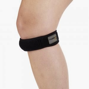
Patellofemoral braces appear to be effective for preventing
anterior knee pain (Yeung et al 2011).

Corrective taping is effective in reducing pain in
patellofemoral pain (kneecap joint) (Herrington & Payton 1997). Tailored
patella taping reduces pain in the short term in patella femoral pain syndrome
(Barton et al 2014).
But…
Taping did not reduce pain in the patellofemoral pain group
but it did enhance the efficiency of vastus medialis oblique (Keet et al 2007).
Acupuncture:
Acupuncture may have a role in the treatment of
tendinopathy, through the facilitation of tendon blood flow and fibroblastic
activity but there is need for further research (Neal & Longbottom 2012).
Injections:
Autologous blood: injected locally to promote repair
activity through the administration of growth factors directly to the site of
injury (Coombes 2010).
However, there is no good quality evidence for autologous
blood injections for chronic tendinopathy when compared to usual care (Rabago
et al 2009).
Platelet-rich plasma: injected locally to promote repair
activity through the administration of concentrated growth factors directly to
the injury site (Coombes 2010).
However, there is insufficient evidence to support the use
of Platelet Rich Therapies for musculoskeletal soft tissue injuries (Moraes et
al 2013).
Aprotinin: injected locally to inhibit collagenase which
would otherwise break down collagen and has been found to be increased in
tendinopathy (Coombes 2010).
Polysulphated glycosaminoglycan: injected locally to prevent
destruction and facilitate repair through inhibiting metalloproteinase enzyme
activity (Coombes 2010).
Botulinum toxin: injected locally to decrease tensile stress
through the tendon and inhibit substance P, which is increased in tendinopathy
(Coombes 2010).
Sodium hyaluronate: injected locally to absorb mechanical
stress and provide a protective buffer for tissues (Coombes 2010).
Corticosteroid: injected locally to down regulate (acting to
decrease) inflammation in the affected tendon (Coombes 2010). Now with this
injection you would definitely not want to do this with a tendinosis and there
is reasoning that maybe it could be detrimental to the tendon in the long term
whether it is tendinosis or tendinitis. This piece of research may not be on
the patella tendon but Tennis elbow is also a tendinopathy:
Corticosteroid injection versus placebo injection resulted
in worse clinical outcomes after 1 year with Tennis elbow (Coombes et al 2013).
Food for thought!
High volume saline: a saline solution is injected along the
surface of the tendon, producing a mechanical effect on the new vascular
ingrowth (Coombes 2010).
Polidocanol: targeted disruption of new vasculature by
administration of a scelerosant to precipitate blood vessel fibrosis (Coombes
2010).
Prolotherapy: hypertonic glucose injected locally to
initiate repair activity by causing local tissue trauma (Coombes 2010).


nice post
ReplyDeletepain physiotherapy
I have read your idea and seen your technique.I think your technique is follow able.I know many technique form your blog. Five Dock Physiotherapist Alina Kennedy
ReplyDeleteNice Post !
ReplyDeleteOrthopedic Hospital in Dombivli || Orthopedic Surgeon in Dombivli || Multispeciality Hospitals in Dombivli || Physiotherapy in Dombivli